Eye Care Services
At Andrew Watkins Optometrist, our highly qualified, experienced Optometrists provide the most advanced eyecare diagnosis and specialised treatment for complex contact lens needs and paediatric vision conditions.
Diabetic retinopathy
Diabetic retinopathy is a complication of diabetes that affects the retina, the light-sensitive tissue at the back of the eye. It can cause vision loss, and in some cases, blindness.
There are two main types of diabetic retinopathy: non-proliferative diabetic retinopathy (NPDR) and proliferative diabetic retinopathy (PDR).NPDR is the milder form of diabetic retinopathy. It is characterized by the growth of small blood vessels in the retina. These blood vessels can leak fluid, causing swelling of the retina.
PDR is the more advanced form of diabetic retinopathy. It is characterized by the growth of new blood vessels in the retina. These blood vessels are fragile and can bleed easily. Bleeding in the retina can cause vision loss.
There are a number of treatments available for diabetic retinopathy. The type of treatment that is best for you will depend on the severity of your retinopathy.Laser treatment is a common treatment for NPDR. Laser treatment can help to seal off leaky blood vessels and prevent them from growing.
Injections of medications called anti-VEGF drugs can also be used to treat NPDR and PDR. These medications help to shrink new blood vessels and prevent them from leaking.Vitrectomy is a surgical procedure that can be used to remove blood from the retina or to remove scar tissue that is causing vision loss.
If you have diabetes, it is important to have regular eye exams. Early detection and treatment of diabetic retinopathy can help to prevent vision loss.Childrens Vision- Myopia/VT/Amblyopia/Strabismus
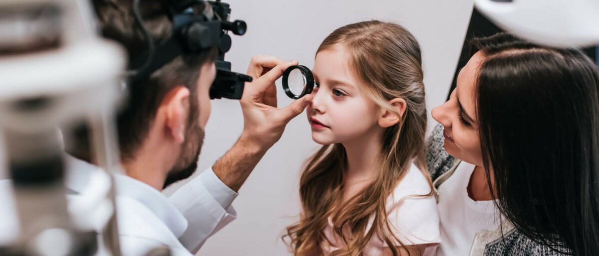
If your child has 20/20 or 6/6 vision, this is only a small part of having good vision. Your child must also be focussed in each eye, must have good eye movement control, good eye-hand co-ordination, good eye health and normal visual perception.
At Andrew Watkins Optometrist we have a special interest in making sure that your child develops the best possible vision. We test all the visual skills necessary develop good vision.
There are many vision problems that children can have. They include:
- Strabismus
- Amblyopia
- Myopia
- Hyperopia
- Astigmatism
- Muscle Inco-ordination
- Colour Vision Defects
- Visual Perceptual Deficits
Some of these are very obvious and are picked up early in childhood and others are much less obvious.
Strabismus is a condition that interferes with binocular vision because it prevents a person from directing both eyes simultaneously to align with each other at the same spot. This is often known as a squint. Strabismus is present in about 4% of children. Treatment should be started as early as possible to ensure the development of the best possible vision.
Amblyopia (also called lazy eye) is a disorder of sight. It results in decreased vision in an eye that otherwise appears normal. Whenever the brain does not receive visual signals from an eye for a long period of time, there is a risk of amblyopia. It also can occur when the brain “turns off” the visual processing of one eye to prevent double-vision. It is common in children with strabismus.
Detecting the condition in early childhood increases the chance of successful treatment; this disorder has been estimated to affect 1–5% of the population.
Myopia is a condition of the eye where the light that comes in does not directly focus on the retina but in front of it, causing the image that one sees when looking at a distant object to be out of focus, but in focus when looking at a close object. It also known as short-sightedness.
Hyperopia is a defect of vision causing difficulty focusing on near objects, and in extreme cases causing a sufferer to be unable to focus on objects at any distance. Children with hyperopia can experience blurred vision, headaches, accommodative dysfunction, binocular dysfunction, amblyopia, and strabismus. Most school age children are in fact slightly hyperopic and therefore must exert an extra effort to bring their vision into sharp focus for both far and near tasks. For some children it will interfere with their ability to do schoolwork.
Astigmatism is a refraction error of the eye in which there is a difference in degree of power in different meridians. Astigmatism causes difficulties in seeing fine detail. Astigmatism can be often corrected by glasses with a lens that corrects for the difference in power.
Muscle Inco-ordination occurs when the complex muscle system for co-ordinating the two eyes to work as a team are not properly balanced. They often occur together with other vision problems and if left untreated contribute to a worsening of the vision problem.
Colour Vision Defects occur in about 9% of boys and 0.5% of girls. They are almost always inherited but can be the result of disease or injury. Almost all people with colour vision defects see most colours but due to the imbalance of their colour receptors they see them slightly differently to the way someone with normal vision sees them. They will therefore have difficulty in identifying some colours and will confuse some colours.
Visual Perception is the ability to analyse and understand what the eyes are seeing. Children with vision problems are more likely to have difficulty with their visual perception; however these problems can occur with otherwise normal vision. If this problem does exist, the underlying vision problem is treated first and then a program of visual perceptual therapy is administered.
Macular Degeneration
Macular degeneration is a condition that causes progressive damage to the macular, the light sensitive tissue at the back of the eye. Macular degeneration is the leading cause of blindness in Australia and will affect 1 in 7 people over the age of 50 and the incidence increases with age*. Those with early macular degeneration may have no noticeable symptoms but the disease can cause central vision loss if not treated early.
Early detection of macular degeneration is aided by having regular eye tests. At Andrew Watkins Optometrist we utilise Optical Coherence Tomography (OCT), a non-invasive imaging test to detect macular degeneration. OCT uses light waves to take cross-sectional images of your retina. OCT allows the Optometrist to see each of the retina’s distinctive layers and pick up early signs of macular degeneration, which can include fatty deposits known as drusen, pigment cell disruption or leaking blood or fluid.
For optimum eye health, it’s recommended that everyone over the age of 40 have their eyes tested every two years.
* Source: Macular Disease Foundation
Optical Coherence Tomography
Optical coherence tomography (OCT) is a non-invasive imaging technique that uses light waves to create detailed images of the retina, the light-sensitive tissue at the back of the eye. OCT can be used to diagnose and monitor a variety of eye conditions, including:
- Age-related macular degeneration (AMD)
- Diabetic retinopathy
- Glaucoma
- Cataracts
- Retinal detachment
OCT can also be used to measure the thickness of the retina and to assess the health of the optic nerve. This information can be used to track the progression of eye diseases and to monitor the effectiveness of treatment.
OCT treatment is typically done in a practice. The patient will sit in a chair and rest their head on a chin rest. The Optometrist will place a contact lens on the patient's eye and then use the OCT machine to scan the retina. The scan takes a few minutes and is painless.
The images created by OCT are stored in a computer and can be reviewed. We can use the images to diagnose eye diseases, to track the progression of eye diseases, and to monitor the effectiveness of treatment.
OCT is a safe and effective imaging technique. There are no known side effects associated with OCT treatment.
If you are concerned about your eye health, talk to our Optometrist about OCT treatment. OCT can be a valuable tool for diagnosing and monitoring eye diseases.
Optos Retinal Imaging
Optos retinal imaging is a non-invasive imaging technique that uses a handheld scanner to capture a 200-degree image of the retina. This allows eye care professionals to view a wider area of the retina than traditional imaging techniques, which can help to detect eye diseases earlier and more accurately.
Optos retinal imaging is used to diagnose and monitor a wide range of eye conditions, including:
- Age-related macular degeneration (AMD)
- Diabetic retinopathy
- Glaucoma
- Retinal detachment
- Uveitis
- Coats' disease
Optos retinal imaging is also used to monitor the progression of eye diseases and to assess the effectiveness of treatment.
The procedure is safe and painless, and it is typically done as part of a routine eye exam. The scanner is held close to the eye, and a flash of light is used to capture the image. The entire procedure takes just a few minutes.
The images captured by Optos retinal imaging are stored in a computer and can be reviewed by the eye care professional. This allows the Optometrist to see a detailed view of the retina and to diagnose any potential problems.
Therapeutics
Therapeutic treatments for eye conditions can include:
Medications: Eye drops, ointments, or oral medications may be prescribed to treat infections, inflammation, allergies, glaucoma, dry eye syndrome, or other eye-related conditions.
Laser Therapy: Laser treatment may be used for various purposes, such as correcting refractive errors (LASIK or PRK), treating glaucoma, or managing certain retinal conditions.
Intraocular Injections: In some cases, medications are directly injected into the eye to treat conditions like macular degeneration, diabetic retinopathy, or retinal vein occlusion.
Surgical Interventions: Therapeutic eye surgeries are performed to address specific eye conditions, such as cataract surgery to remove a cloudy lens, corneal transplant surgery to treat certain corneal disorders, or retinal surgery to repair retinal detachments or tears.
Therapeutic Contact Lenses: Specially designed contact lenses may be used for therapeutic purposes, such as managing corneal irregularities or promoting healing in certain eye conditions.
The specific therapeutic approach depends on the diagnosis and the underlying eye condition being treated. It is important to consult with an optometrist, to determine the most appropriate therapeutic treatment for an individual's specific eye condition.
Sports Vision
Sports Vision: Improve Your Performance on the Field
Sports vision is a field of optometry that focuses on improving visual performance in sports. Sports vision therapy can help athletes of all ages and skill levels improve their eye-hand coordination, depth perception, visual tracking, and other visual skills that are important for success in sports.
Why is sports vision important?
Good sports vision can give you an edge in competition. It can help you:
- See the ball more clearly and track it better
- Make faster and more accurate decisions
- React more quickly to changes in the environment
- Stay focused and avoid getting distracted
- How can sports vision therapy help?
Sports vision therapy is a customized program of exercises and activities that are designed to improve your visual skills. The exercises are tailored to your individual needs and goals, and they can be done at home or in the office of an optometrist or vision therapist.
What are the benefits of sports vision therapy?
Sports vision therapy can help you improve your:
- Eye-hand coordination
- Depth perception
- Visual tracking
- Visual reaction time
- Visual focus
- Visual memory
- Visual attention
Who can benefit from sports vision therapy?
Sports vision therapy can benefit athletes of all ages and skill levels, from recreational players to professional athletes. It can also be helpful for people who participate in activities that require good visual skills, such as driving, golfing, and hunting.
If you are an athlete who is looking to improve your performance, talk to our Optometrist about sports vision therapy. They can assess your visual skills and recommend a program that is right for you.
Contact Lenses
Many spectacle wearers spend years unaware that they are suitable for contact lenses. At Andrew Watkins Optometrist, our qualified Optometrists will fit you with the most appropriate contact lenses for your prescription and lifestyle.
Contact lenses are available to correct both distance, near and multifocal prescriptions, as well as for patients with astigmatism. We will help you find the right contact lens to suit your specific needs.
Eye Examination
Our Optometrists play an important role in your eye health.
If you have not had your eyes examined for quite some time you may not be aware of all the tests involved in a comprehensive eye examination. If it has been more than two years since your last eye test, we generally allow 45 minutes to complete a comprehensive consultation.
Our team of experienced Optometrists detect, diagnose, and treat eye health and vision conditions that affect vision including glaucoma, macular degeneration, diabetic retinopathy, hypertension, lazy eye and cataracts.
They can also identify general health conditions that are first detected via an eye exam, provide referrals to eye surgeons (ophthalmologists) and often help manage post-eye-surgery health.
The majority of our services attract a Medicare rebate.
Every consultation and eye examination is with an experienced Optometrist using the latest and most advanced eyecare procedures. With our full eye health examination (45 minutes), comprehensive internal and external eye testing is standard and includes:
- Field of vision
- Eye muscle control
- Visual acuity
- Ability to focus
- Ability to see colour
We request you bring the following to your eye examination
- Your medicare card
- Your latest pair of prescription glasses
- Your latest pair of prescription sunglasses
- Any contact lenses you may use
- Previous prescription details or Optometrist’s reports if you are new to our practice
Orthokeratology
Orthokeratology (ortho-k) is the fitting of specially designed gas permeable contact lenses (RGP) that you wear overnight. While you are asleep, the lenses gently reshape the front surface of your eye (cornea) so you can see clearly the following day after you remove the lenses when you wake up. Ortho-k lenses are prescribed for two purposes: To correct refractive errors To slow the progression of childhood myopia
Eye Care Services
At Andrew Watkins Optometrist, our highly qualified, experienced Optometrists provide the most advanced eyecare diagnosis and specialised treatment for complex contact lens needs and paediatric vision conditions.
Bringing new hope to sufferers of Dry Eye syndrome, IPL Treatment (Intense Pulsed Light) is now available at Insight Optometrists, Indooroopilly.
Dry Eye syndrome is an irritating condition, ranging from general eye discomfort to severe inflammation of the upper eyelid. It is aggravated by our modern life with many factors contributing to Dry Eyes: artificial lighting, screen work, air conditioning, pollution and even contact lenses.
Previously, there was no cure for Dry Eyes, only symptomatic treatments like eye drops, artificial tears, steam glasses or manual stimulation of the Meibomian glands.
After IPL treatment, immediate improvement is usually seen and, in as few as three sessions, substantial results can be obtained to reduce the effect of dry eyes*. Symptoms are reduced and the health and moisture of the eyes is restored, bringing comfort and relief from this painful condition for the longer term.
As a rapid, effective and affordable alternative to previous treatments, IPL uses pulsating waves of light to stimulate the malfunctioning Meibomian glands of the eye. It is these glands that, when healthy, secrete the external lipid layer of the lacrimal film that provides lubrication of the eye’s surface and prevents evaporation of the tear film.
Source: Studies by E-Swin https://www.e-swin.com/medical-research/meibomian-blepharitis/
What does IPL treatment involve?
With the patient seated in a reclining chair, eye goggles are worn to protect the eyes. The eye area is cleaned and a protective gel applied to the skin below the eyes. Five flashes of light are applied under the lower eyelid on each eye. The process only takes a few minutes to complete.
Is it safe?
IPL has been used in the cosmetic industry for 20 years, and the light flashed in the Dry Eye treatment is at less than half the strength than that used in, for example, pigmentation treatment. There is no risk of skin cancer from IPL because the light is filtered to ensure it is well outside dangerous limits. The treatment is not suitable for pregnant women.
Free Dry Evaluation
IPL treatment is suitable for most Dry Eye sufferers, however, Insight Optometrists offer a Free Dry Eye Evaluation to assess your condition and plan a personalised treatment program at no obligation. This consultation will confirm the best course of action to bring relief from your symptoms and will include the use of IPL and other treatments where appropriate.
Orthokeratology (ortho-k) is the fitting of specially designed gas permeable contact lenses (RGP) that you wear overnight. While you are asleep, the lenses gently reshape the front surface of your eye (cornea) so you can see clearly the following day after you remove the lenses when you wake up. Ortho-k lenses are prescribed for two purposes: To correct refractive errors To slow the progression of childhood myopia
Diabetic retinopathy is a complication of diabetes that affects the retina, the light-sensitive tissue at the back of the eye. It can cause vision loss, and in some cases, blindness.
There are two main types of diabetic retinopathy: non-proliferative diabetic retinopathy (NPDR) and proliferative diabetic retinopathy (PDR).
NPDR is the milder form of diabetic retinopathy. It is characterized by the growth of small blood vessels in the retina. These blood vessels can leak fluid, causing swelling of the retina.
PDR is the more advanced form of diabetic retinopathy. It is characterized by the growth of new blood vessels in the retina. These blood vessels are fragile and can bleed easily. Bleeding in the retina can cause vision loss.
There are a number of treatments available for diabetic retinopathy. The type of treatment that is best for you will depend on the severity of your retinopathy.
Laser treatment is a common treatment for NPDR. Laser treatment can help to seal off leaky blood vessels and prevent them from growing.
Injections of medications called anti-VEGF drugs can also be used to treat NPDR and PDR. These medications help to shrink new blood vessels and prevent them from leaking.
Vitrectomy is a surgical procedure that can be used to remove blood from the retina or to remove scar tissue that is causing vision loss.
If you have diabetes, it is important to have regular eye exams. Early detection and treatment of diabetic retinopathy can help to prevent vision loss.
If your child has 20/20 or 6/6 vision, this is only a small part of having good vision. Your child must also be focussed in each eye, must have good eye movement control, good eye-hand co-ordination, good eye health and normal visual perception.
At xxx we have a special interest in making sure that your child develops the best possible vision. We test all the visual skills necessary develop good vision.
There are many vision problems that children can have. They include:
- Strabismus
- Amblyopia
- Myopia
- Hyperopia
- Astigmatism
- Muscle Inco-ordination
- Colour Vision Defects
- Visual Perceptual Deficits
Some of these are very obvious and are picked up early in childhood and others are much less obvious.
Strabismus is a condition that interferes with binocular vision because it prevents a person from directing both eyes simultaneously to align with each other at the same spot. This is often known as a squint. Strabismus is present in about 4% of children. Treatment should be started as early as possible to ensure the development of the best possible vision.
Amblyopia (also called lazy eye) is a disorder of sight. It results in decreased vision in an eye that otherwise appears normal. Whenever the brain does not receive visual signals from an eye for a long period of time, there is a risk of amblyopia. It also can occur when the brain “turns off” the visual processing of one eye to prevent double-vision. It is common in children with strabismus.
Detecting the condition in early childhood increases the chance of successful treatment; this disorder has been estimated to affect 1–5% of the population.
Myopia is a condition of the eye where the light that comes in does not directly focus on the retina but in front of it, causing the image that one sees when looking at a distant object to be out of focus, but in focus when looking at a close object. It also known as short-sightedness.
Hyperopia is a defect of vision causing difficulty focusing on near objects, and in extreme cases causing a sufferer to be unable to focus on objects at any distance. Children with hyperopia can experience blurred vision, headaches, accommodative dysfunction, binocular dysfunction, amblyopia, and strabismus. Most school age children are in fact slightly hyperopic and therefore must exert an extra effort to bring their vision into sharp focus for both far and near tasks. For some children it will interfere with their ability to do schoolwork.
Astigmatism is a refraction error of the eye in which there is a difference in degree of power in different meridians. Astigmatism causes difficulties in seeing fine detail. Astigmatism can be often corrected by glasses with a lens that corrects for the difference in power.
Muscle Inco-ordination occurs when the complex muscle system for co-ordinating the two eyes to work as a team are not properly balanced. They often occur together with other vision problems and if left untreated contribute to a worsening of the vision problem.
Colour Vision Defects occur in about 9% of boys and 0.5% of girls. They are almost always inherited but can be the result of disease or injury. Almost all people with colour vision defects see most colours but due to the imbalance of their colour receptors they see them slightly differently to the way someone with normal vision sees them. They will therefore have difficulty in identifying some colours and will confuse some colours.
Visual Perception is the ability to analyse and understand what the eyes are seeing. Children with vision problems are more likely to have difficulty with their visual perception; however these problems can occur with otherwise normal vision. If this problem does exist, the underlying vision problem is treated first and then a program of visual perceptual therapy is administered.
Behavioural eye conditions are caused by problems with the way the brain processes visual information. They can be caused by a variety of factors, including genetics, learning disabilities, and brain injuries.
The treatment for behavioural eye conditions will vary depending on the specific condition. However, some common treatments include:
Vision therapy (VT): This is a type of therapy that helps to improve the way the brain processes visual information.
- Occupational therapy (OT): This is a type of therapy that helps to improve the coordination of the eyes and the hands.
- Prism glasses: These glasses can help to align the eyes or to improve convergence.Surgery: Surgery may be an option for some people with strabismus or DVD.
Macular degeneration is a condition that causes progressive damage to the macular, the light sensitive tissue at the back of the eye. Macular degeneration is the leading cause of blindness in Australia and will affect 1 in 7 people over the age of 50 and the incidence increases with age*. Those with early macular degeneration may have no noticeable symptoms but the disease can cause central vision loss if not treated early.
Early detection of macular degeneration is aided by having regular eye tests. At Davis Eyecare we utilise Optical Coherence Tomography (OCT), a non-invasive imaging test to detect macular degeneration. OCT uses light waves to take cross-sectional images of your retina. OCT allows the Optometrist to see each of the retina’s distinctive layers and pick up early signs of macular degeneration, which can include fatty deposits known as drusen, pigment cell disruption or leaking blood or fluid.
For optimum eye health, it’s recommended that everyone over the age of 40 have their eyes tested every two years.
*Source: Macular Disease Foundation
Optical coherence tomography (OCT) is a non-invasive imaging technique that uses light waves to create detailed images of the retina, the light-sensitive tissue at the back of the eye. OCT can be used to diagnose and monitor a variety of eye conditions, including:
- Age-related macular degeneration (AMD)
- Diabetic retinopathy
- Glaucoma
- Cataracts
- Retinal detachment
OCT can also be used to measure the thickness of the retina and to assess the health of the optic nerve. This information can be used to track the progression of eye diseases and to monitor the effectiveness of treatment.
OCT treatment is typically done in a practice. The patient will sit in a chair and rest their head on a chin rest. The Optometrist will place a contact lens on the patient’s eye and then use the OCT machine to scan the retina. The scan takes a few minutes and is painless.
The images created by OCT are stored in a computer and can be reviewed. We can use the images to diagnose eye diseases, to track the progression of eye diseases, and to monitor the effectiveness of treatment.
OCT is a safe and effective imaging technique. There are no known side effects associated with OCT treatment.
If you are concerned about your eye health, talk to our Optometrist about OCT treatment. OCT can be a valuable tool for diagnosing and monitoring eye diseases.
Optos retinal imaging is a non-invasive imaging technique that uses a handheld scanner to capture a 200-degree image of the retina. This allows eye care professionals to view a wider area of the retina than traditional imaging techniques, which can help to detect eye diseases earlier and more accurately.
Optos retinal imaging is used to diagnose and monitor a wide range of eye conditions, including:
- Age-related macular degeneration (AMD)
- Diabetic retinopathy
- Glaucoma
- Retinal detachment
- Uveitis
- Coats’ disease
Optos retinal imaging is also used to monitor the progression of eye diseases and to assess the effectiveness of treatment.
The procedure is safe and painless, and it is typically done as part of a routine eye exam. The scanner is held close to the eye, and a flash of light is used to capture the image. The entire procedure takes just a few minutes.
The images captured by Optos retinal imaging are stored in a computer and can be reviewed by the eye care professional. This allows the Optometrist to see a detailed view of the retina and to diagnose any potential problems.
Therapeutic treatments for eye conditions can include:
Medications: Eye drops, ointments, or oral medications may be prescribed to treat infections, inflammation, allergies, glaucoma, dry eye syndrome, or other eye-related conditions.
Laser Therapy: Laser treatment may be used for various purposes, such as correcting refractive errors (LASIK or PRK), treating glaucoma, or managing certain retinal conditions.
Intraocular Injections: In some cases, medications are directly injected into the eye to treat conditions like macular degeneration, diabetic retinopathy, or retinal vein occlusion.
Surgical Interventions: Therapeutic eye surgeries are performed to address specific eye conditions, such as cataract surgery to remove a cloudy lens, corneal transplant surgery to treat certain corneal disorders, or retinal surgery to repair retinal detachments or tears.
Therapeutic Contact Lenses: Specially designed contact lenses may be used for therapeutic purposes, such as managing corneal irregularities or promoting healing in certain eye conditions.
The specific therapeutic approach depends on the diagnosis and the underlying eye condition being treated. It is important to consult with an optometrist, to determine the most appropriate therapeutic treatment for an individual’s specific eye condition.
Sports Vision: Improve Your Performance on the Field
Sports vision is a field of optometry that focuses on improving visual performance in sports. Sports vision therapy can help athletes of all ages and skill levels improve their eye-hand coordination, depth perception, visual tracking, and other visual skills that are important for success in sports.
Why is sports vision important?
Good sports vision can give you an edge in competition. It can help you:
- See the ball more clearly and track it better
- Make faster and more accurate decisions
- React more quickly to changes in the environment
- Stay focused and avoid getting distracted
- How can sports vision therapy help?
Sports vision therapy is a customized program of exercises and activities that are designed to improve your visual skills. The exercises are tailored to your individual needs and goals, and they can be done at home or in the office of an optometrist or vision therapist.
What are the benefits of sports vision therapy?
Sports vision therapy can help you improve your:
- Eye-hand coordination
- Depth perception
- Visual tracking
- Visual reaction time
- Visual focus
- Visual memory
- Visual attention
Who can benefit from sports vision therapy?
Sports vision therapy can benefit athletes of all ages and skill levels, from recreational players to professional athletes. It can also be helpful for people who participate in activities that require good visual skills, such as driving, golfing, and hunting.
If you are an athlete who is looking to improve your performance, talk to our Optometrist about sports vision therapy. They can assess your visual skills and recommend a program that is right for you.
Many spectacle wearers spend years unaware that they are suitable for contact lenses. At Practice namexx, our qualified Optometrists will fit you with the most appropriate contact lenses for your prescription and lifestyle.
Contact lenses are available to correct both distance, near and multifocal prescriptions, as well as for patients with astigmatism. We will help you find the right contact lens to suit your specific needs.